Electrophysiologic study (EPS)
The derivation of electrical signals from the heart with special catheters has lost much of its importance in the pure diagnosis of cardiac arrhythmias. Less than 5% of the catheter-based electrophysiological procedures we perform are purely diagnostic. However, EPU is still a prerequisite and thus an integral part of catheter ablation for triggering and characterising a cardiac arrhythmia.
Diagnostic EPS still occasionally plays a role in securing the indication for pacemaker implantation and in characterising the risk of sudden cardiac death.
Under local anaesthesia and general sedation ("sleep anaesthesia"), accesses (locks) are placed via a blood vessel in the groin region, through which one to two (rarely several) thin catheters are advanced into the heart. The catheters enable the conduction of electrical signals from different heart cavities with a high precision in the range of a few milliseconds. Through electrical stimulation, the physiological and pathophysiological conduction properties of the heart muscle and the specific conduction system can be characterised and any cardiac arrhythmias can be triggered or terminated. Often, the triggering of arrhythmias can be additionally influenced by the administration of short-acting drugs.
A diagnostic EPU rarely takes longer than 20 to 45 minutes and is - even if it may look different at first glance - a procedure with very few complications.
If the aim is to clarify cardiovascular concomitant diseases, which plays a decisive role in the assessment of cardiac arrhythmias, further examinations are possible in addition to pure rhythm diagnostics at Swiss Ablation: these primarily include ultrasound procedures, ergometry and laboratory analyses.
Elektrocardiogramm (ECG)
is the recording of all electrical activities of the heart muscle cells by means of an electrocardiograph. You could also call it the individual handwriting of the heart. Adhesive electrodes attached to the surface of the body can perceive the voltage changes of these electrical activities absolutely painlessly and record them over time. This results in the recurring image of a heart action. In the electrophysiological examination, the ECG is not obtained from the surface but directly from inside the heart.
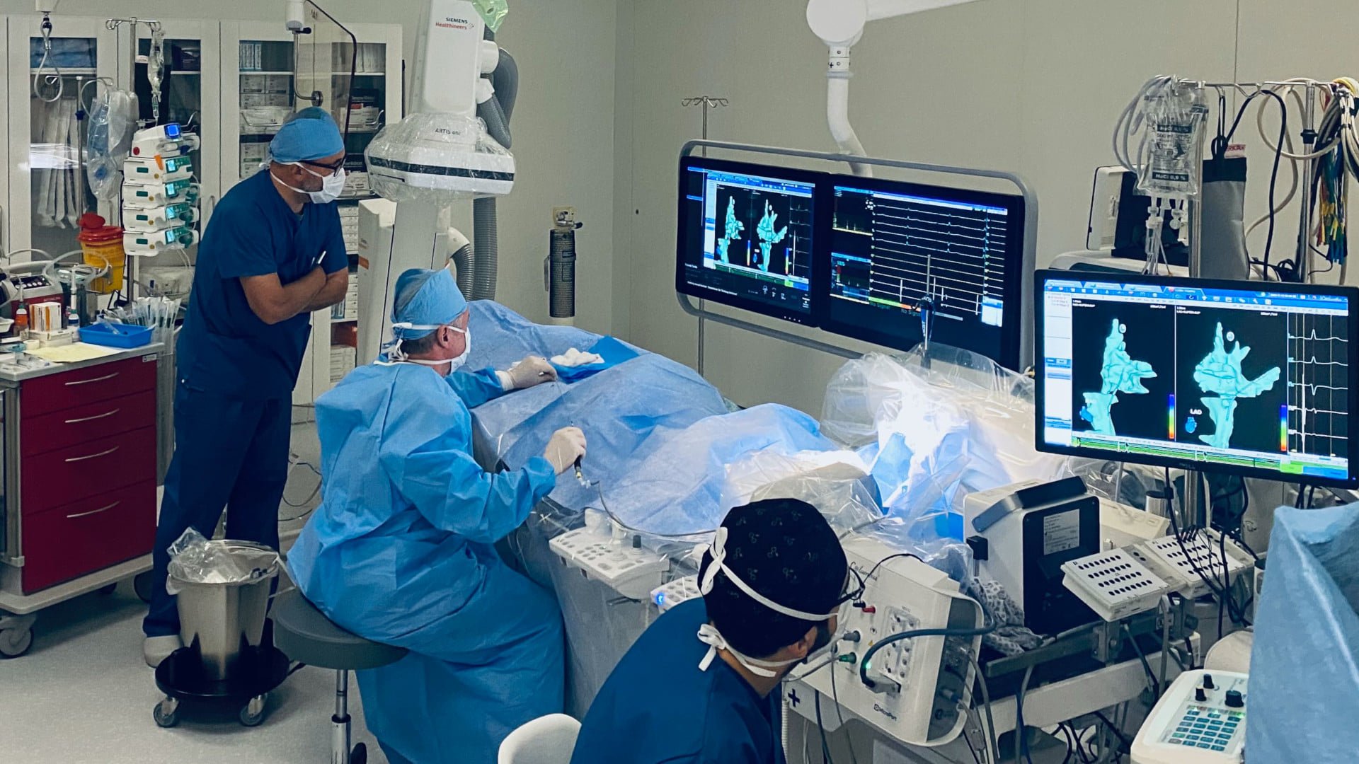
3D Mapping and catheter ablation
In this specially equipped operating theatre, work is carried out in a sterile environment and with an X-ray system. The catheters can be located by X-rays, but recently also by electromagnetic fields (without radiation). These catheters are then inserted into the heart via the inguinal vein and have a locating or stimulating function (diagnostic catheters for mapping or stimulation of the heart) or they are catheters that transfer energy to certain regions of the heart with the aim of destroying the tissue there (ablation catheters).
Implantable loop recorder (ILR)
Implantable event recorders are i-chips that are placed under the skin under local anaesthesia. With these recordings, which are stored digitally every day, the ECG can be recorded continuously. This allows us to clarify all forms of cardiac arrhythmia, especially in patients with unknown cardiac arrhythmia or symptoms such as dizziness and syncope.